Knee Muscle Anatomy Mri / Figure 14 from Normal MR imaging anatomy of the knee. | Semantic Scholar. The knee joint is the junction of the thigh and leg. Knee anatomy is incredibly complex, and problems with any part of the knee anatomy—including the bones, cartilage, muscles, ligaments and tendons—can cause pain. Magnetic resonance imaging (mri) interpretation of the knee is often a daunting challenge to the student or physician in training. Rubin da, kettering jm, towers jd, britton ca: Want to learn more about it?
On anatomical parts the user. Master leg and knee anatomy using our topic page. Normal knee mri for reference. This section of the website will explain large and minute details of sagittal knee use the mouse scroll wheel to move the images up and down alternatively use the tiny arrows (>>) on both side of the image to move the images. Knee anatomy francesc malagelada jordi vega pau golanó the knee is the largest joint in the human body and one of the most complex from a functional point of view.
MRI shoulder anatomy | shoulder coronal anatomy | free cross sectional anatomy from mrimaster.com These muscles work in groups to flex, extend and stabilize the extending along the anterior surface of the thigh are the four muscles of the quadriceps femoris group (vastus lateralis, vastus medialis, vastus. Each anatomical structure was labeled interactively. It is a noninvasive test that can visualize the inner structures of the knee, including the cartilage and ligaments, the surface of the bones, and the muscles and tendons that surround the knee joint. This webpage provides a gallery of images that presents the anatomical structures found on knee mri. The muscles of the knee include the quadriceps, hamstrings, and the muscles of the calf. Master leg and knee anatomy using our topic page. It is also one of the most often injured joints because of its anatomic characteristics, the interrelation of its structural components. Free cross sectional anatomy of the knee based on mri :
Injuries of the patellofemoral joint.
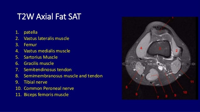
General anatomy and musculoskeletal system. Please email baodo at stanford.edu. Mri for evaluating knee pain in older patients: Knee anatomy francesc malagelada jordi vega pau golanó the knee is the largest joint in the human body and one of the most complex from a functional point of view. The knee is designed to fulfill a number of functions: Quadriceps tendon semitendinosus tendonsemimembranosus muscle popliteal artery and vein biceps femoris femur vastus medialis sartorius muscle suprapatellar bursa. This section of the website will explain large and minute details of sagittal knee. Learn about mri anatomy with free interactive flashcards. Tendons attach the muscles to each other. Normal knee mri for reference. Scroll through the structures to understand the anatomy. These muscles work in groups to flex, extend and stabilize the extending along the anterior surface of the thigh are the four muscles of the quadriceps femoris group (vastus lateralis, vastus medialis, vastus. Injuries of the patellofemoral joint.
The knee is designed to fulfill a number of functions: Mri patterns of neuromuscular disease involvement thigh & other muscles 2. Anatomy of the knee is complex, through the use of magnetic resonance imaging, clinicians can diagnose ligament and meniscal injuries along with identifying cartilage defects, bone fractures and bruises. Knowing about knee anatomy can help people understand how knee arthritis develops and sometimes causes pain. Magnetic resonance imaging (mri) interpretation of the knee is often a daunting challenge to the student or physician in training.
MRI anatomy of the knee www.unidadortopedia.com PBX: 6923370. Unidad Especializada en Ortopedia ... from i.pinimg.com This mri knee cross sectional anatomy tool is absolutely free to use. Injuries of the patellofemoral joint. Involved early gray = muscle: The muscles that affect the knee's movement run along the thigh and calf. Knowing about knee anatomy can help people understand how knee arthritis develops and sometimes causes pain. Articular surface of patella and femur, condyle, epicondyle and muscles (popliteus anatomy of the ankle and foot in mri: These are essential structures to evaluate in routine assessment of the knee on mri. Scroll through the structures to understand the anatomy.
They are attached to the femur (thighbone), tibia (shinbone), and fibula (calf bone) by fibrous tissues called ligaments.
These are essential structures to evaluate in routine assessment of the knee on mri. The muscles that affect the knee's movement run along the thigh and calf. Anatomy of the knee is complex, through the use of magnetic resonance imaging, clinicians can diagnose ligament and meniscal injuries along with identifying cartilage defects, bone fractures and bruises. Learn about mri anatomy with free interactive flashcards. The knee joint is the junction of the thigh and leg. It is a noninvasive test that can visualize the inner structures of the knee, including the cartilage and ligaments, the surface of the bones, and the muscles and tendons that surround the knee joint. View of the anatomical labels. Use the checklist to quiz yourself. Rubin da, kettering jm, towers jd, britton ca: Want to learn more about it? They are attached to the femur (thighbone), tibia (shinbone), and fibula (calf bone) by fibrous tissues called ligaments. Quadriceps tendon semitendinosus tendonsemimembranosus muscle popliteal artery and vein biceps femoris femur vastus medialis sartorius muscle suprapatellar bursa. Each anatomical structure was labeled interactively.
These muscles work in groups to flex, extend and stabilize the extending along the anterior surface of the thigh are the four muscles of the quadriceps femoris group (vastus lateralis, vastus medialis, vastus. Each anatomical structure was labeled interactively. Knee anatomy francesc malagelada jordi vega pau golanó the knee is the largest joint in the human body and one of the most complex from a functional point of view. On anatomical parts the user. Learn about the muscles, tendons, bones, and ligaments that comprise the knee joint anatomy.
Knee Mri Anatomy - Anatomy Drawing Diagram from image.slidesharecdn.com Involved early gray = muscle: This webpage provides a gallery of images that presents the anatomical structures found on knee mri. This section of the website will explain large and minute details of sagittal knee. Tendons attach the muscles to each other. The knee joint is most significantly affected by two major muscle groups: See the pictures and anatomy description of knee joint bones, cartilage, ligaments, muscle and tendons with resources for knee problems & injuries. This mri knee cross sectional anatomy tool is absolutely free to use. It is a noninvasive test that can visualize the inner structures of the knee, including the cartilage and ligaments, the surface of the bones, and the muscles and tendons that surround the knee joint.
Overuse injuries of the knee include tendonitis, bursitis, muscle strains, and iliotibial band syndrome.
Free cross sectional anatomy of the knee based on mri : Technical considerations for mri evaluation of the knee extensor mechanism. Use the checklist to quiz yourself. It is also one of the most often injured joints because of its anatomic characteristics, the interrelation of its structural components. This section of the website will explain. Normal knee mri for reference. Mri for evaluating knee pain in older patients: The muscles of the knee include the quadriceps, hamstrings, and the muscles of the calf. Knowing about knee anatomy can help people understand how knee arthritis develops and sometimes causes pain. Helps to lower and raise the body. Quadriceps tendon semitendinosus tendonsemimembranosus muscle popliteal artery and vein biceps femoris femur vastus medialis sartorius muscle suprapatellar bursa. Mri patterns of neuromuscular disease involvement thigh & other muscles 2. Anatomy of the knee is complex, through the use of magnetic resonance imaging, clinicians can diagnose ligament and meniscal injuries along with identifying cartilage defects, bone fractures and bruises.
Share :
Post a Comment
for "Knee Muscle Anatomy Mri / Figure 14 from Normal MR imaging anatomy of the knee. | Semantic Scholar"


Post a Comment for "Knee Muscle Anatomy Mri / Figure 14 from Normal MR imaging anatomy of the knee. | Semantic Scholar"